Color Doppler ultrasound is a technique that enables the physician to identify the direction and speed of blood flow within a vessel or heart chamber. Color Doppler is not part of the Standard Examination of the heart, but is used by a specialist with special training in fetal echocardiography. The use of color Doppler ultrasound for evaluation of the fetal heart and detection of birth defects was first reported by Dr. DeVore in 1987. Since this first publication over 200 articles have been published in the medical literature describing the use of this technology for evaluating the fetal heart. Color Doppler displays the flow of blood based upon its direction of flow; red-orange towards to top of the transducer, blue away from the transducer. The following is an example of this concept.
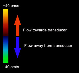
This illustrates the color bars and direction of flow. The transducer is always located at the top of the image. Therefore, when blood flow is towards the transducer it is depicted in red, and away from the transducer in blue. The different hues of red-orange and blue-green indicate the different velocities or speed that blood is flowing. For example, if the color Doppler display is yellow then it is flowing towards the transducer faster than if it is deep red.
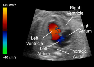
This illustrates the use of color Doppler to identify the flow of blood into the ventricles. In this example blood flow is only observed flowing into the left ventricle, but not the right ventricle. Therefore, the right ventricle is underdeveloped. The blue demonstrates blood flowing between the right atrium and the left atrium.
The Use of Color Doppler to Detect Fetal Heart Abnormalities
The examples that follow illustrate the use of color Doppler to either diagnose a heart abnormality,or enhance the diagnosis when compared to 2D imaging.
Ebstein's Anomaly
This is the 2D image of the four-chamber view. The right atrium is enlarged with what appears to be an abnormal tricuspid valve.
The color Doppler defines the underlying pathology by demonstrating abnormal blood flow back into the right atrium from the right ventricle. The abnormal flow originates from a displaced tricuspid valve which is located lower in the ventricle than it should be. This is called Ebstein's malformation often seen in women who take anti-depressants such as Lithium.
This is the labeled image of the pathology demonstrating abnormal blood flow back into the right atrium from the right ventricle. The abnormal flow originates within the right ventricle. RA=right atrium, LA=left atrium, RV=right ventricle, LV=left ventricle, TR=tricuspid regurgitation.
Hypoplastic Right Ventricle
This is the 2D image of the four-chamber view. Because of the age of the fetus, the anatomy of the four-chamber view is difficult to evaluate.
The color Doppler defines the underlying pathology by demonstrating the flow patterns within the heart.
This is the labeled image of the pathology demonstrating several features. When the heart fills with blood (diastole) blood is observed filling only the left ventricle. When the heart begins to contract (systole) blood is observed going across the ventricular septal defect to fill the smaller right ventricle. The arrows illustrate the ventricular septal defect (VSD). RA=right atrium, LA=left atrium, RV=right ventricle, LV=left ventricle.
Tricuspid Regurgitation
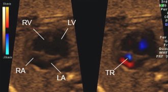
This image illustrates the four-chamber view using 2D ultrasound on the left and color Doppler on the right. Tricuspid regurgitation cannot be demonstrated using 2D ultrasound. This finding is important because tricuspid regurgitation is associated with an increased risk for Down syndrome when it is observed during the first or second trimeters of pregnancy.
This image illustrates the four-chamber view using 2D ultrasound on the left and color Doppler on the right. Tricuspid regurgitation cannot be demonstrated using 2D ultrasound. This finding is important because tricuspid regurgitation is associated with an increased risk for Down syndrome when it is observed during the first or second trimeters of pregnancy.RA=right atrium, LA=left atrium, RV=right ventricle, LV=left ventricle.
Ventricular Septal Defect
This image illustrates the four-chamber view using 2D ultrasound. While a ventricular septal defect is present, it is difficult to recognize from the 2D image.
This illustrates a shunting ventricular septal defect located in the lower septum. The color Doppler clearly shows this defect that was not apparent on the 2D study.
This is the labeled image of the pathology demonstrating the shunting ventricular septal defect (VSD) easily identified with color Doppler ultrasound RA=right atrium, LA=left atrium, RV=right ventricle, LV=left ventricle.